© Copyright 2002 by Paul Ramp. All rights reserved.
Chapter 40: Development
Objectives:
- describe and identify the structure of the sperm and egg when they are ready for fertilization
- describe the events of fertilization and mechanisms to prevent multiple fertilizations
- describe the changes to a fertilized egg through implantation in the uterus and formation of the placenta
- describe the development of the embryo through the first two months of the pregnancy
- describe the changes in the fetus that occur in the last 7 months of development
- describe the processes that lead to the birth of the child
From this unit you should be able to:
Web Text:
- - Overview -
- - Structure of the Sperm and Egg -
- - Fertilization -
- - Preembryonic Period -
- - Embryonic Period -
- - Fetal Period -
- - Birth -
Overview - In this unit we'll be looking at some of the events that occur between the time of the unfertilized egg within the fallopian tube and the birth of a child. These events are occurring during the gestation period, defined as the time between the end of the last menstrual cycle and the birth of the child. The gestation period is quite variable in mammals. In humans the gestation period is 280 days on average. In terms of the actual development time, recall that release and potential fertilization of the egg does not occur until about 14 or 15 days into the menstrual cycle so the actual development time from fertilization to birth is about 266 days.
There are many ways one can divide the gestation period into smaller time periods. The most common is to simply divide these 280 days or 9 months based on the calendar. Consequently we may divide the pregnancy into three, thee month trimesters. While dividing the pregnancy into trimesters is easy and convenient, such a subdivision is arbitrary from a biological point of view. Here, after discussing the events of fertilization, we will divide the pregnancy into three periods that are marked by biological events occurring with the developing child. These three periods are the peembryonic period, the embryonic period and the fetal period.
Structure of the Sperm and Egg - Before looking into the events of fertilization, let's take a closer look at the cells involved in this event, the sperm and the egg or oocyte.
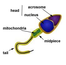
| The body of a sperm cell can be divided into three regions, the head, a midpiece and a tail. The head is something of a teardrop shaped structure with the cell's nucleus located at the wider base. At the tip of this structure is an organelle called the acrosome. This is a vesicle that contains hydrolytic enzymes, enzymes that are digestive. The midpiece of the sperm lies behind the head. It is a like a short rod in shape. Within the midpiece are many mitochondria. Mitochondria are cellular organelles that make molecules of ATP, the molecules that are used as an energy source by the cell. The final section of the sperm is the tail. The tail consists of a long flagellum or axial filament running down its center. Surrounding the axial filament for most of its length is a sheath made from fibrous proteins. The tail, using the energy from the midpiece, rapidly moves back and forth to propel the sperm forward. |
Structure of a sperm cell |
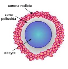
| After release from the ovary the oocyte is surrounded by layers of granulosa cells from the follicle. This layer of cells surrounding the oocyte is called the corona radiata or jelly coat. While these cells serve to protect the egg after it is released from the ovary, they also create a barrier the sperm must penetrate. Between the corona radiata and the oocyte is a thick extracellular membrane that has been secreted by the granulose cells. This is the zona pellucida. Inside the oocyte the nucleus is resting at the end of meiosis I, waiting for fertilization to signal its completion of meiosis II. |
Structure of a secondary oocyte |
Fertilization - When reaching the ovum and contacting the corona radiata, the sperm release the enzymes contained within the acrosome. These enzymes begin to digest the cells of the corona radiata, allowing the sperm to push their way down through these cells. The quantity of enzymes release from a single sperm is not sufficient to all that sperm to penetrate completely through this barrier. The sperm that arrive later will find the corona radiata already weakened by the actions of the earlier sperm and these later sperm will have a better chance of successfully breaking through this barrier.
The cells of the corona radiata provide one mechanism for maintaining the integrity of the species. Species specific proteins mark both the sperm and egg and only sperm with the correct protein markers are able to penetrate this barrier.
Once a sperm has penetrated the corona radiata, it move through the zona pellucida and makes contact with the egg's plasma membrane. The membrane of the sperm and egg unite, sending the sperm's nucleus into the egg's cytoplasm.
The moment the egg and sperm membrane's unite, several processes are triggered that will prevent a second sperm from fertilizing with the egg. The first of these is a rapid, temporary change to the egg's membrane called the fast block response. When the sperm unites with the oocyte, calcium ions are rapidly released from the oocyte resulting it a change in the egg's membrane potential, similar to the change in membrane potential that occurs with a nervous impulse. With is altered membrane potential, additional sperm are unable to unite with the egg. The change in membrane potential also triggers other events in the egg. Meiosis of the egg's nucleus resumes so that it can quickly complete meiosis II so that the egg's genetic material can unite with the sperm's. The change in membrane potential also triggers organelles called cortical granules to migrate to the plasma membrane and release their contents into the extracellular space of the zona pellucida.
Of course the egg's membrane can not remain depolarized for a prolonged period of time. That would severely disrupt cellular activities needed to permit the cell to survive and grow. To permanently block additional sperm from fertilizing the egg a second mechanism, the slow block response is utilized. The substances released into the zona pellucida by the cortical granules alter the chemical nature of this layer and alters its osmotic potential. Essentially, these changes make the zona pellucida impenetrable by sperm. The change in osmotic potential causes water to enter this area, expanding this extracellular membrane. This altered zona pellucida is called the fertilization membrane.
In humans, about 20 minutes after the sperm and egg unite, meiosis II is completed. The nuclei of the sperm and egg unite and fertilization is completed. Immediately the cell, now a zygote, begins to divide. This marks the beginning of the fist stage of development, the preembryonic period.
Preembryonic Period - The preembryonic period of development last for approximately two week. During this time, the developing embryo is passing down the fallopian tube and entering the uterus.
The earliest event of the preembryonic period is called cleavage. After the first cleavage, the embryo consists of two cells. Then these divide to produce four cell, then eight, 16, 32 and so on. While these cells are dividing there is little cell growth. Consequently this developing ball of cells remains about the same size as the egg cell at the time of fertilization. These cells form a solid mass called a morula.
By about the fourth or fifth day after fertilization, the cell mass consists of about 100 or so cells. At this time the cells are pushed away from each other to from a hollow sphere-like structure called a blastocyst. Just a single layer of cells makes up the wall of the blastocyst. This cell layer is called to trophoblast. It is destined to develop into part of the placenta. On one side of the blastocyst, just inside the tophoblast, is a mass of cells called the inner cell mass. Only a few of the cells within this inner cell mass are destined to develop into the actual offspring.
The second event of the preembryonic period occurs about the 6th or 7th day after fertilization. This is implantation in which the blastocyst embeds itself into the endometrial wall of the uterus. When the blastocyst contacts the uterine wall, the trophoblast is stimulated to rapidly divide and secrete an enzyme which dissolves the endometrium. This allows the blastocyst to bury into this tissue. The endometrium quickly heals over the point of entry, sealing the blastocyst within the endometrium.
Marking the end of the preembryonic phase is placentation, the development of the placenta. The cells of the trophoblast, now called the chorion, have digested the surrounding cells of the uterus to create a pool of blood containing nutrients that can be absorbed by the developing embryo. These cells of the developing embryo that have specialized to exchange nutrients and wastes make up the placenta. As the placenta develops, its cells begin to secrete increasing quantities of estrogens and progesterone. These hormones prevent the next menstrual cycle from starting. If the placenta does not release enough of these hormones, menstruation will begin as usual and the pregnancy will be aborted.
Embryonic Period - The embryonic period extends from about week 3 to week 8 after fertilization. Two major events are completed during this period. These are the formation of the extraembryonic membranes and gastrulation.
|
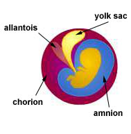
The extraembryonic membranes are tissues derived from the developing embryo that produce sac-like structures that help support embryonic and fetal development. Few traces of these remain in the adult organism. There are four extraembryonic membranes. These are the amnion, the yolk sac, the allantosis and the chorion. The amnion is a membrane formed from cells that eventually completely surround the developing embryo. This creates a sac around the embryo that becomes filled with a watery fluid, the amniotic fluid. The amniotic fluid provides a favorable environment around the embryo. During an amniocentesis, a sample of this fluid is taken. Since the embryo continually sheds cells into this fluid, these cells may be examined for the presence of some birth defects. |
Extraembryonic membranes |
As the name implies, nutrient rich yolk is stored in the yolk sac. This structure is quite large in organisms that produce large quantities of yolk but in humans the yolk sac is relatively small. Although small, the yolk sac still has important contributions to the development of the embryo. This is the site where the first blood cells of the embryo are produced. Blood vessels are first formed here.
First forming as an out pocketing at the base of the yolk sac is the allantosis. In egg laying organisms, the allantosis serves as a site for the storage of metabolic wastes. In humans, the allantosis makes up a part of the umbilical cord, connecting the embryo with the placenta. The base of the allantosis develops into part of the urinary bladder of the adult organism.
The fourth extraembryonic membrane is the chorion. This is formed from the trophoblast and from cells of the inner cell mass that have spread around the inner surface of the trophoblast. The chorion forms the fetal portion of the placenta, allowing the exchange of nutrients and wastes between the mother and developing child. Since the cells of the chorion are genetically identical to the fetus, collecting some of these cells by a procedure called chorionic villi sampling (CVS) can be used to look for some birth defects.
The second major event that is completed early in the embryonic phase is gastrulation. This is a process of cell migrations that occur within the inner cell mass that give rise to three primary layers of tissue called germ layers. These three layers are the ectoderm on the outside of the developing embryo, the endoderm on the inside of the developing embryo and the mesoderm between the ectoderm and endoderm.
The outer germ layer, the ectoderm, gives rise to the skin covering the body. It also gives rise to the cornea and lens of the eye, the epithelium of the mouth and nasal cavities, tooth enamel, the epithelium of some glands such as part of the pituitary and the adrenal medulla, as well as some of the bones of the face. In addition the ectoderm gives rise to all of the body's nervous tissue. This occurs shortly after gastrulation when two ridges of tissue along the dorsal side fold together, fusing to form the dorsal hollow nerve cord. The anterior portion of this neural tube enlarges to become the brain.
The middle germ layer, the mesoderm, gives rise to all of the muscle tissue in the body. Also derived from the mesoderm are cartilage, bone, blood and other connective tissues. The epithelium of the blood vessels and lymphatic system as well as the membrane of joint cavities are derived from mesoderm. Most of the organs of the urogenital system, the kidneys, ureters, gonads and reproductive ducts, are derived from the mesoderm.
The endoderm, the inner most germ layer, gives rise to the digestive tract including the liver and the pancreas. The epithelium of the respiratory tract is derived from endoderm. Also arising from endoderm tissue are the thyroid, parathyroid and thymus. The lower portion of the urogenital system, the urethra and bladder, are derived from endoderm tissue.
The embryonic period of development ends at about two months. At this time, the embryo is approximately 28 mm in length and weighs about 2.7 grams. By this time all organ systems have been initiated and are forming. The basic structure of the heart has developed and has been beating since about 22 days after fertilization. The digestive tract is formed and its different subdivisions are becoming specialized. The thymus, thyroid, pituitary and adrenal glands have formed. Ossification of bones is just beginning and the skeletal muscles of the distinct limbs are forming.


